Francis Crick, Rosalind Franklin, James Watson, and Maurice Wilkins
These four scientists codiscovered the double-helix structure of DNA, which formed the basis for modern biotechnology.
Scientists from many countries, representing several scientific disciplines, were trying to understand the mysteries of life and heredity in the decades around 1900. Looking at everything from grasshoppers to bacteria, they made new knowledge about the chemistry of living cells and the transmission of hereditary information. By the 1950s, many scientists were convinced that the particles carrying this information from one generation to the next were made of deoxyribonucleic acid (DNA), and several of them began trying to unravel its chemical structure. Using a variety of different methods, Francis Crick (1916–2004), Rosalind Franklin (1920–1958), James Watson (1928–2025), and Maurice Wilkins (1916–2004) contributed to the 1953 announcement that DNA was a double helix.
As the Nobel Prize commission later recognized, knowledge of the double helix held immense “significance for information transfer in living material.” In other words, understanding the structure of the molecule helped to explain how it could copy itself, passing on instructions from one generation to the next.
DNA, the “Transforming Principle”
What are genes made of? In the 1930s, most scientists would have said proteins. This began to change in 1944, when researchers at the Rockefeller Institute for Medical Research demonstrated that it was DNA—not proteins or ribonucleic acid (RNA)—that caused bacteria to transform. The research was published in a paper for the Journal of Experimental Medicine, adding to a huge amount of interest in the hereditary material of genes and viruses.
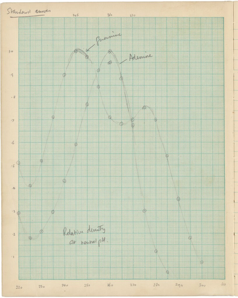
New experimental techniques and imaging processes were used to further study DNA. These revealed it to be a large molecule made up of sugar, phosphate, and an additional four compounds—adenine (A), cytosine (C), guanine (G), and thymine (T). Extending this work, biochemist Erwin Chargaff identified an interesting feature of DNA base pairs, noting that A and T tended to occur in roughly equal amounts, as did C and G. These discoveries were foundational to the later work on the structure of DNA.
Competition and Collaboration
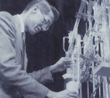
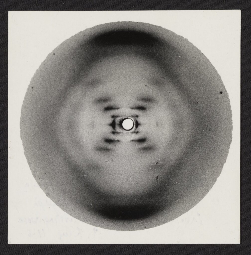
Maurice Wilkins and his student Raymond Gosling began studying DNA with X-ray diffraction crystallography at King’s College London (KCL) around 1950. Developed by William Henry Bragg and his son, Lawrence, the technique helps scientists see the structure of crystallized material. Beams of X-radiation are aimed at the crystal, bending as they pass through it. The positions and intensities of these “diffracted” X-rays are then recorded on film for analysis. The technique was fairly new to Wilkins and Gosling when they obtained an especially pure sample of DNA from chemist Rudolf Signer in 1950. Prepared from calf thymus (probably from Signer’s local butcher shop), the Signer DNA could be stretched into very thin samples, which yielded remarkably clear X-ray diffraction patterns in turn.
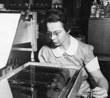
Biophysicist John T. Randall, who oversaw the laboratory where Wilkins and Gosling worked, was eager to recruit someone with more experience in X-ray diffraction. Randall was relatively open-minded when it came to women in science (8 of the 31 researchers in his unit were women, a high percentage at the time). In 1951, he offered a position to Rosalind Franklin, a physical chemist with expertise in the X-ray analysis of coal. With her arrival, responsibility for various subjects and techniques became confused, creating intense rivalries within Randall’s lab.
Franklin Before DNA
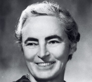
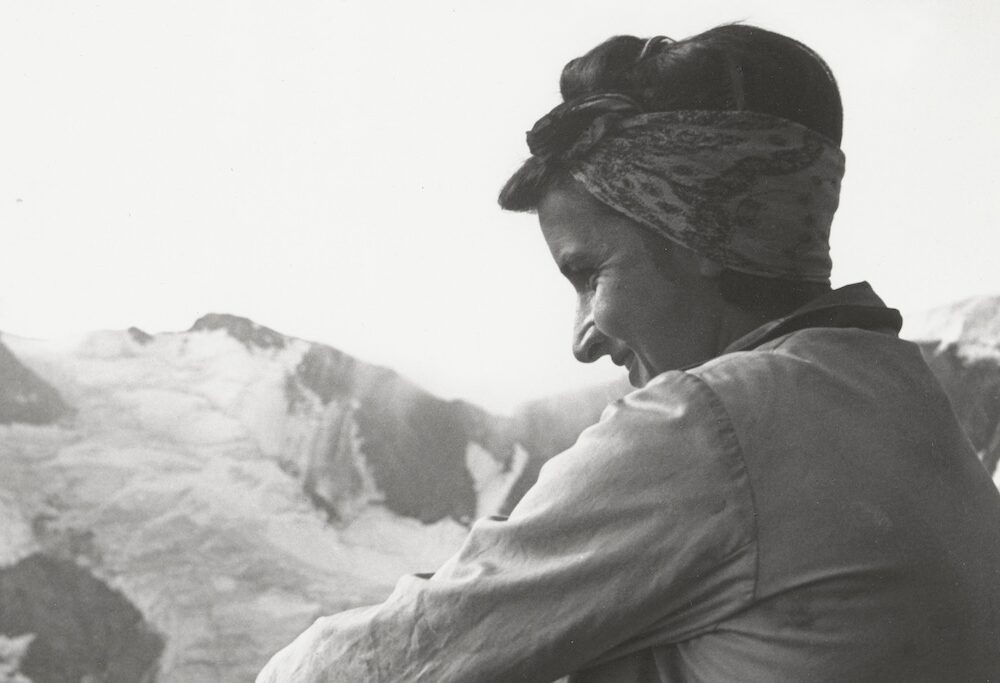
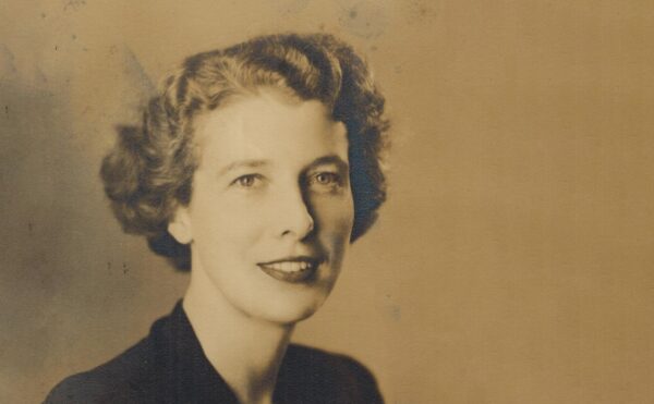
Rosalind Franklin was born into a prominent Jewish family in London on July 25, 1920. Her parents were committed to education as a means of promoting social and personal well-being. As a child, she assisted her father, Ellis, in campaigns to support the Working Men’s College, which offered evening classes to laborers who would not otherwise have had the opportunity to study. Simultaneously Franklin was embarking on her own elite education. After early graduation from St. Paul’s Girls’ School, she attended Newnham College, the oldest women’s college at Cambridge University, where she specialized in physical chemistry.
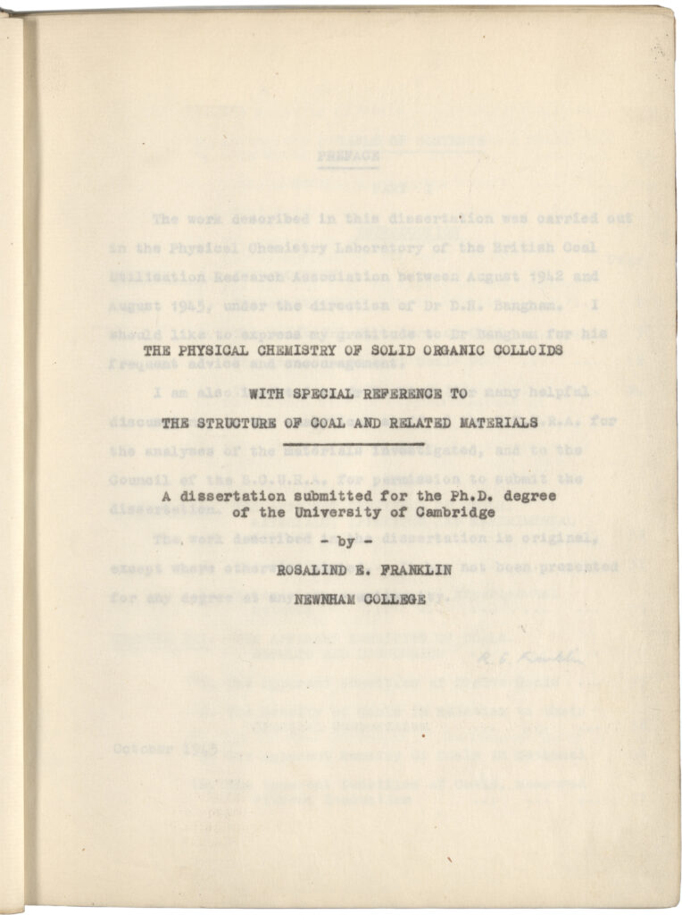
During World War II, Franklin was torn between a strong personal desire to take on war work, and her dedication to scientific research. It came as a relief when she secured a position at the British Coal Utilization Research Association (BCURA). There, she performed fundamental investigations on the properties of coal and graphite. This work became the basis of her PhD dissertation in 1945 and the focus of her first publications.
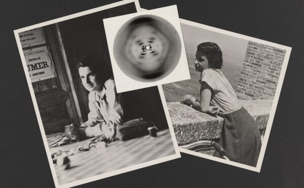
After the war, Franklin obtained an appointment at the Laboratoire Centrale des Services Chimiques de l’Etat in Paris. She gained rigorous training in X-ray crystallography through this postdoctoral position and rapidly became a respected authority in the field. As thoroughly as she enjoyed her time in Paris, by 1951 she felt pulled to return to England. She applied for a fellowship and received funding to support work in Randall’s biophysics unit at KCL that year.
From Physics to Biology
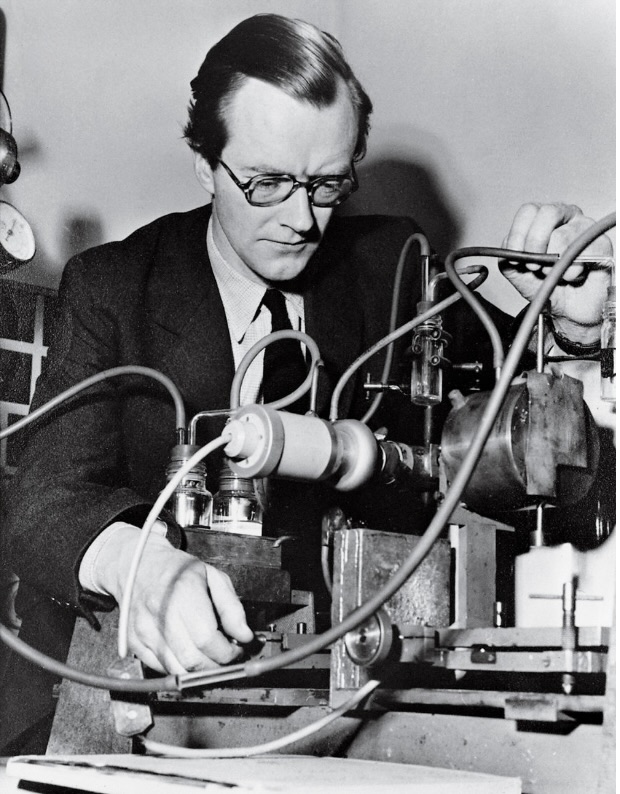
Maurice Wilkins was Randall’s second-in-command at the KCL biophysics unit. Born in Pongaroa, a small New Zealand farming town, Wilkins traveled to England as a young boy to begin his education. He earned an undergraduate degree in physics from St. John’s College, Cambridge, in 1938, and his PhD from Birmingham University two years later. In this early work, Wilkins studied the way solids emit light and trap heat. This research proved strategic during World War II: Wilkins applied his theoretical knowledge of phosphorescence to the improvement of cathode-ray tube screens used for radar systems. Around the same time, he began working on the separation of uranium isotopes for use in bombs. He joined the Manhattan Project in 1944.
Like many other nuclear physicists, Wilkins became disillusioned with his research when he saw the destruction brought by atomic bombs in 1945. This propelled him towards new interdisciplinary projects in biophysics. He and Randall—who had undergone a similar conversion—established labs for biophysical studies first at the University of St. Andrews in Scotland and then at KCL. Wilkins applied himself to a wide range of biophysical projects. Between 1946 and 1950, for instance, he worked to improve the reflecting microscopes used to study nucleic acids in cells; he also worked on the orientation of purines and pyrimidines in Tobacco Mosaic Virus (TMV). After obtaining the Signer DNA in 1950, Wilkins collaborated closely with Gosling on X-ray diffraction studies. Together they were able to create a perfectly crystalline sample of DNA by manipulating its moisture content. This made it possible to improve upon earlier X-ray studies that had produced blurry images.
Base Pairs
In stories about the discovery of DNA, theoretical modelling sometimes overshadows the physical and experimental work of the biophysicists at KCL. The stereochemistry and modelling of DNA are particularly associated with James Watson and Francis Crick, both of whom were working at Cambridge University’s Cavendish Laboratory in the early 1950s. Watson also shared their side of the story in his controversial 1968 book, The Double Helix: A Personal Account of the Discovery of the Structure of DNA. The book characterized this episode in the history of molecular biology as a race to the finish, and it was particularly unkind to Franklin.
James Watson had traveled a long way to Cambridge. Born on the South Side of Chicago in 1928, he was interested in birds from a young age. He secured one scholarship to study zoology at the University of Chicago, and another for his PhD studies at Indiana University. His dissertation was an investigation of the ways in which bacteriophages (the viruses that invade bacteria in order to reproduce) respond to high energy X-rays.
Watson went to Denmark for postdoctoral research upon his graduation in 1950. His wanted to continue studying viruses and to improve his understanding of chemistry. At a 1951 conference held at the Zoological Station in Naples, he heard Wilkins speak on the molecular structure of DNA and saw some of the X-ray images from the King’s group. This motivated Watson to shift his research program toward the structural chemistry of nucleic acids and proteins. He moved to the Cavendish Laboratory in October 1951 to pursue these interests.
Soon after, Watson met Francis Crick. Crick had earned a bachelor’s degree in physics from University College London and had started graduate work towards a PhD when World War II broke out. Unlike Franklin, who managed to unite her war work with her research interests, an appointment with the British Admiralty drew Crick away from his doctoral studies. After several years working on underwater explosives, Crick left the naval service to study biology in 1947. Like Wilkins, he was eager after the war to turn from instruments of death to the study of life.
Crick was extremely capable, but lacked a background in the life sciences. He spent his first few years at the Cavendish Laboratory learning the fundamentals of biology, organic chemistry, and X-ray crystallography. As a student, he collaborated on a general theory of the diffraction patterns caused by helical structures. Similar work was being done at the same time by Linus Pauling on the helical shapes of certain proteins—knowledge that relied on both theoretical models and experimental evidence. Helical shapes were a hot topic: they reconciled the complexity of living systems with elegant simplicity at the molecular level.
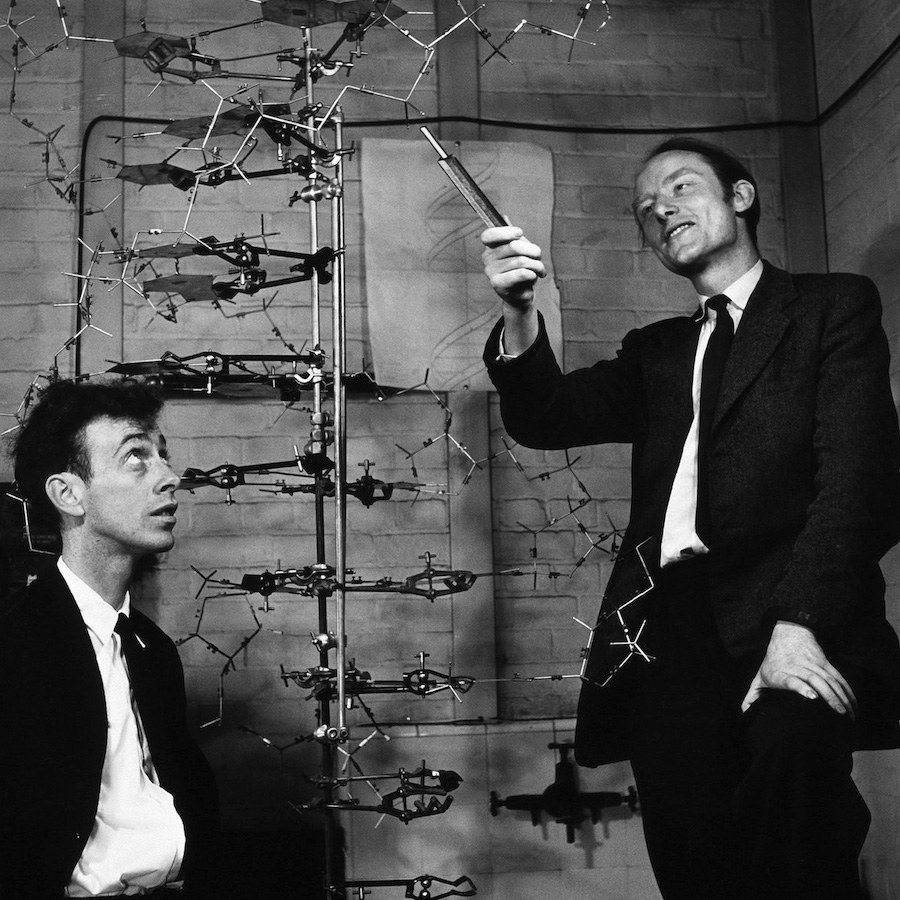
Watson and Crick decided to tackle the problem of DNA structure. With the experimental X-ray diffraction evidence available by 1951, and a growing understanding of the stereochemistry of polynucleotide chains, they felt confident and proposed an initial model toward the end of 1951. It was defined by a three-chain helix with the bases on the outside. But colleagues quickly pointed out this was impossible. Watson and Crick had failed to account for the way the proposed molecule would behave when hydrated: this shape would have completely fallen apart.
It wasn’t until Wilkins showed Watson an especially clear diffraction image taken with a fully hydrated DNA molecule (the so-called “B form”) that Watson and Crick recognized the solution to the problem. This image, known as Photo 51, clearly showed two helices and allowed for very precise mathematical analyses of diffraction intensities. This led Watson and Crick to conclude that the phosphate-sugar chains making up the outside of the twisted ladder ran in opposite directions with the base pairs meeting them perpendicularly at specific distances. In turn, this structure explained how the chains, when pulled apart, could serve as a blueprint for the synthesis of new proteins in the body.
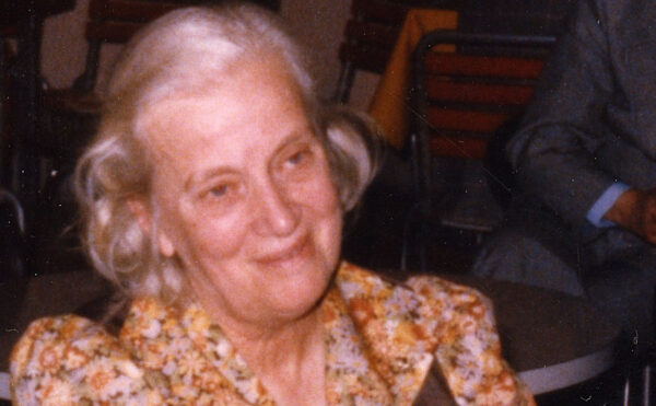
In 1953, Watson and Crick published a short, strongly worded article in Nature announcing the double helix. Nine years later, Watson, Crick, and Wilkins jointly received the Nobel Prize in Physiology or Medicine for their work on the mechanisms of heredity. By tradition, Nobel Prizes were not awarded posthumously at that time. Franklin, who died of ovarian cancer in 1958, was not mentioned in the award.
After the Double Helix
Franklin had not been happy at King’s College, and she left to work in J. D. Bernal’s X-ray crystallography laboratory at Birkbeck College not long after taking Photo 51. She made important contributions to the structural analysis of TMV and Poliovirus at Birkbeck, drawing on some of the experimental techniques she had developed through the study of DNA. The success of Franklin’s “Virus lab” depended on her skillful ability to negotiate access to the best samples, equipment, and experimental data that colleagues had to offer; this included ongoing friendly interactions with both Watson and Crick. She gained public recognition for her work on TMV when Lawrence Bragg invited her to construct a large demonstration model of her findings for the 1958 World’s Fair in Brussels. Sadly, Franklin did not live long enough to read the favorable reviews of her exhibition in the newspapers.
Francis Crick was still a graduate student when his landmark publication with Watson appeared in Nature—he completed his PhD in 1954 with a thesis titled “X-ray Diffraction: Polypeptides and Proteins.” For the next few years, he continued to develop his expertise in X-ray techniques, turning to the study of RNA and protein synthesis at the Brooklyn Polytechnic Institute. Crick went on to investigate the biological significance of the double-helical structure. He articulated the idea that the sequencing of bases functions like “code.” He also identified a process that came to be known as the “central dogma” of molecular biology, the idea that genetic information flows irreversibly in one direction from DNA to RNA to proteins.
Wilkins stayed at King’s College. He applied X-ray methods to the structural determination of nerve cell membranes and RNA. He steadily rose through the ranks at King’s, taking on more responsibility as biophysics grew into a self-standing field of research.
Watson’s later career was more complicated. He joined the Harvard Biology Department in 1956, where he too turned to questions about the role of RNA in protein synthesis. In 1968, he became the director of Cold Spring Harbor Laboratory (CSHL), making it a center of research in molecular biology. From 1988 to 1992, he led the National Center for Human Genome Research at the U.S. National Institutes of Health, becoming the founding director of the Human Genome Project in 1990. Watson’s racist remarks about the intelligence of people of African descent in 2007 prompted the CSHL to revoke his chancellorship. When Watson doubled down on his racist views in a 2019 documentary, his honorary titles were revoked as well. Watson’s fame as a discoverer of the structure of DNA made his continued expressions of sexist, racist, and eugenicist views particularly harmful.
Glossary of Terms
Purines and pyrimidines
Two types of nucleotide bases: guanine and adenine are purines; cytosine and thymine are pyrimidines.
Back >>
Stereochemistry
The three-dimensional study of how atoms are arranged within molecules.
Back >>
Eugenics
Describing various efforts to “improve” specific human populations through activities including selective breeding, immigration restrictions, and sterilization.
Back >>
Further Resources
Consortium for History of Science, Technology and Medicine. Podcast series: The DNA Papers.
Franklin, R. E. & G. G. Gosling. “Molecular Configuration in Sodium Thymonucleate.” Nature 171 (1953): 740–741.
Watson, J. D. & F. H. C. Crick. “Molecular Structure of Nucleic Acids: A Structure for Deoxyribose Nucleic Acid.” Nature 171 (1953): 737–738.
Wilkins, M. H. F., A. R. Stokes & H. R. Wilson. “Molecular Structure of Deoxypentose Nucleic Acids.” Nature 171 (1953): 738–740.
You might also like
SCIENTIFIC BIOGRAPHIES
Florence Ogilvy Bell
What do shark fins, wool, and DNA have in common? Physicist Florence Bell studied them all with X-ray crystallography.
PROJECTS & INITIATIVES
History of Molecular Biology Collection
This unparalleled collection documents the race to identify DNA’s double-helix structure and other significant developments that formed the foundation of molecular biology.
SCIENTIFIC BIOGRAPHIES
Dorothy Crowfoot Hodgkin
Using X-ray crystallography, Hodgkin determined the structures of penicillin, insulin, and vitamin B12 and was the third woman ever to win the Nobel Prize in Chemistry.